Home

Quemar Preparación Pasteles dapi staining concentration Posicionamiento en buscadores Puerto marítimo Contribución

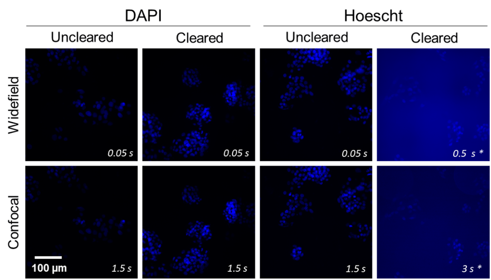
Visikol® - Blog Post: Optimizing staining protocols with Visikol HISTO  clearing: DAPI vs Hoescht | Visikol
Visikol® - Blog Post: Optimizing staining protocols with Visikol HISTO clearing: DAPI vs Hoescht | Visikol

A) DAPI fluorescent staining images of 3T3/NIH fibroblast cell line... |  Download Scientific Diagram
A) DAPI fluorescent staining images of 3T3/NIH fibroblast cell line... | Download Scientific Diagram
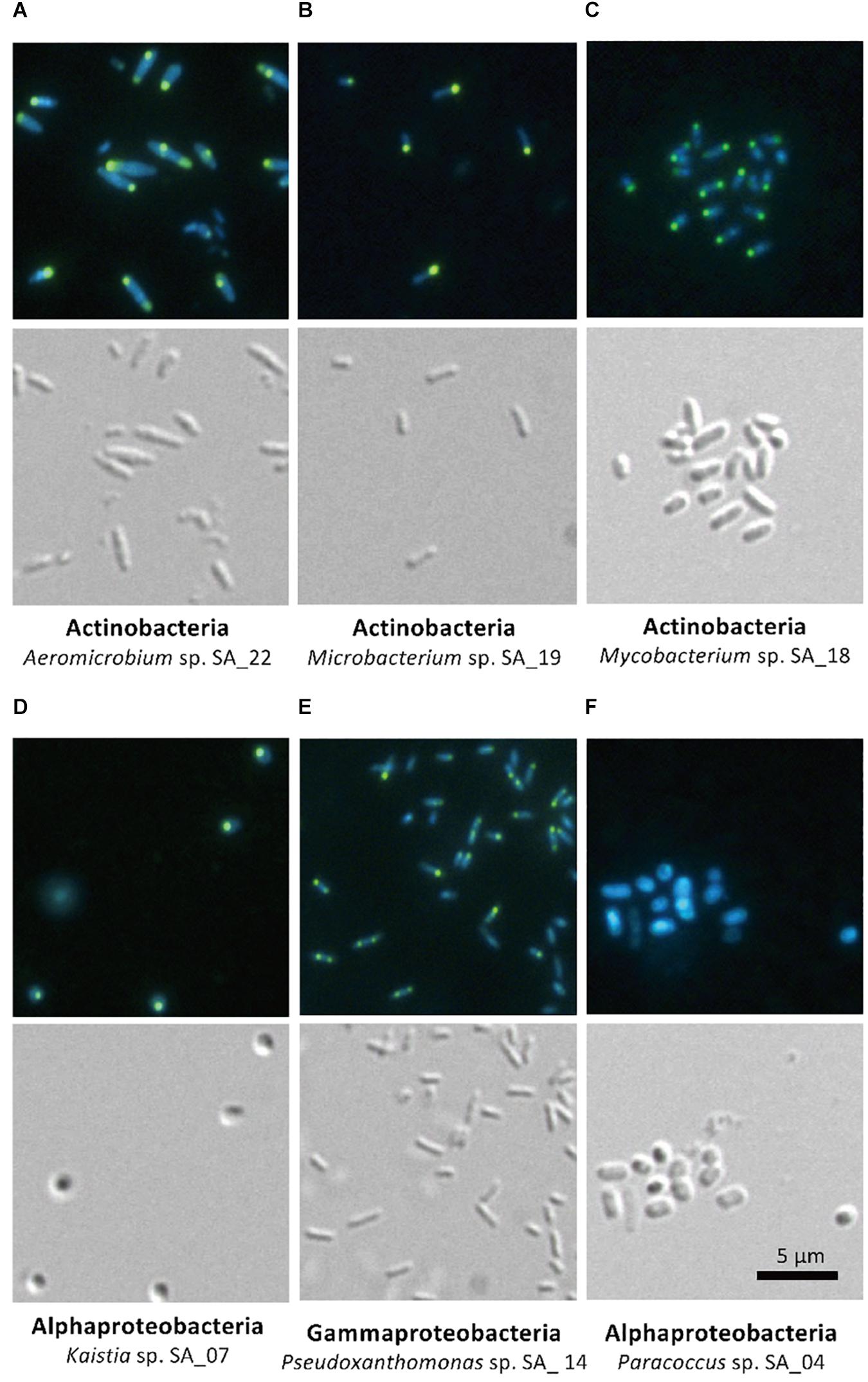
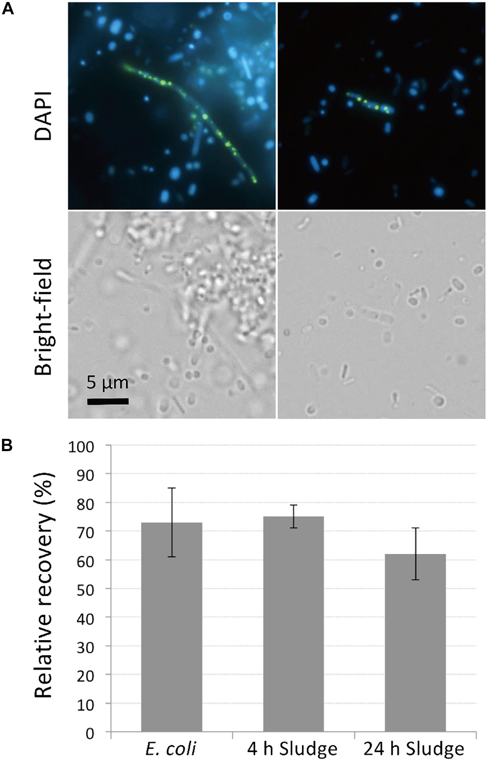
Frontiers | Rapid Enrichment and Isolation of Polyphosphate-Accumulating  Organisms Through 4'6-Diamidino-2-Phenylindole (DAPI) Staining With  Fluorescence-Activated Cell Sorting (FACS)
Frontiers | Rapid Enrichment and Isolation of Polyphosphate-Accumulating Organisms Through 4'6-Diamidino-2-Phenylindole (DAPI) Staining With Fluorescence-Activated Cell Sorting (FACS)
MTT assay, DAPI staining, and flow cytometry of 4T1: (a) 4T1 cells... |  Download Scientific Diagram
MTT assay, DAPI staining, and flow cytometry of 4T1: (a) 4T1 cells... | Download Scientific Diagram

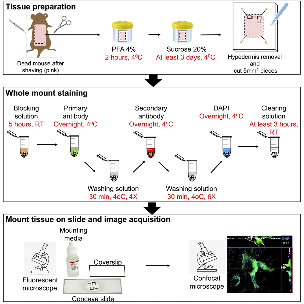
Whole-mount staining coupled to a UV-inducible basal cell carcinoma murine  model
Whole-mount staining coupled to a UV-inducible basal cell carcinoma murine model

Frontiers | Rapid Enrichment and Isolation of Polyphosphate-Accumulating  Organisms Through 4'6-Diamidino-2-Phenylindole (DAPI) Staining With  Fluorescence-Activated Cell Sorting (FACS)
Frontiers | Rapid Enrichment and Isolation of Polyphosphate-Accumulating Organisms Through 4'6-Diamidino-2-Phenylindole (DAPI) Staining With Fluorescence-Activated Cell Sorting (FACS)

A comprehensive toolkit for quick and easy visualization of marker  proteins, protein-protein interactions and cell morphology in Marchantia  polymorpha | bioRxiv
A comprehensive toolkit for quick and easy visualization of marker proteins, protein-protein interactions and cell morphology in Marchantia polymorpha | bioRxiv
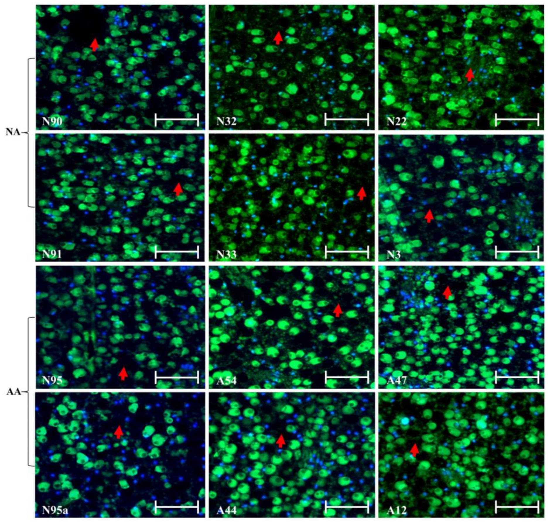
Plants | Free Full-Text | TUNEL Assay and DAPI Staining Revealed Few  Alterations of Cellular Morphology in Naturally and Artificially Aged Seeds  of Cultivated Flax
Plants | Free Full-Text | TUNEL Assay and DAPI Staining Revealed Few Alterations of Cellular Morphology in Naturally and Artificially Aged Seeds of Cultivated Flax

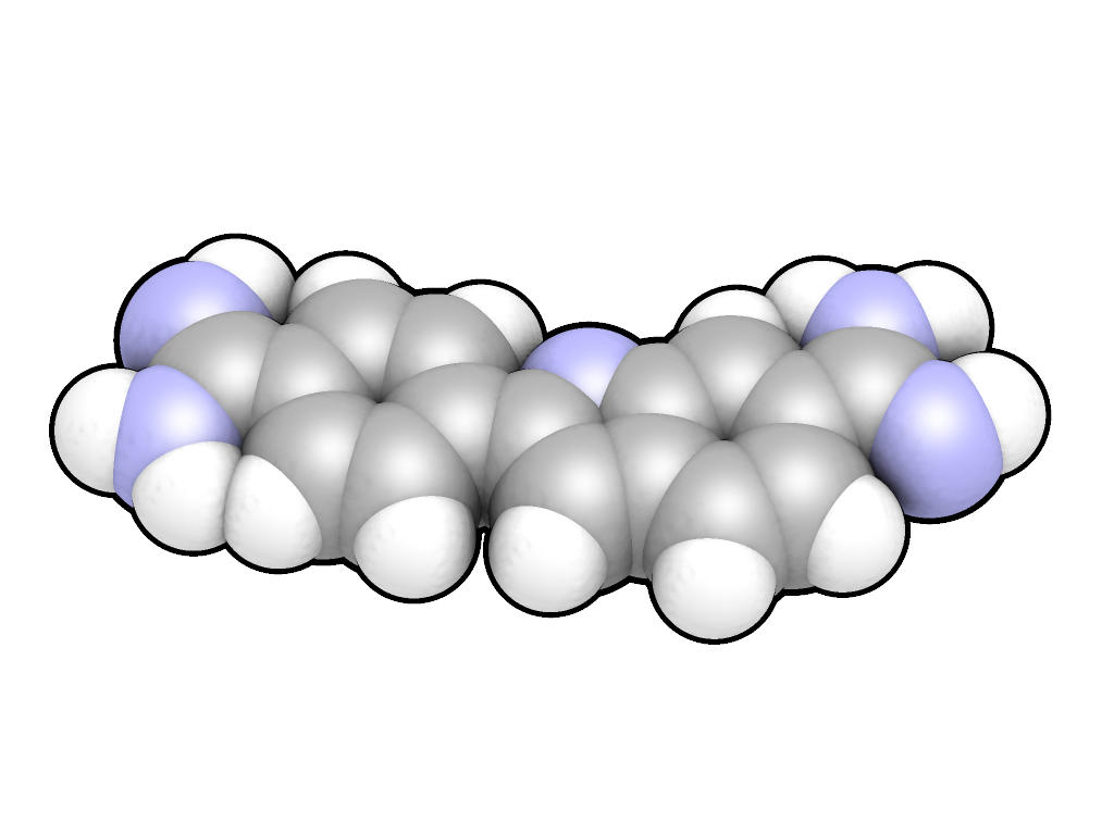
DAPI - Wikipedia
DAPI - Wikipedia

Differential Attentional Process Inventory - an overview | ScienceDirect  Topics
Differential Attentional Process Inventory - an overview | ScienceDirect Topics

Apoptosis detection by DAPI staining. HT-29 cells were treated with... |  Download Scientific Diagram
Apoptosis detection by DAPI staining. HT-29 cells were treated with... | Download Scientific Diagram

Detection of nuclear fragmentation in A549 cells by DAPI staining after...  | Download Scientific Diagram
Detection of nuclear fragmentation in A549 cells by DAPI staining after... | Download Scientific Diagram
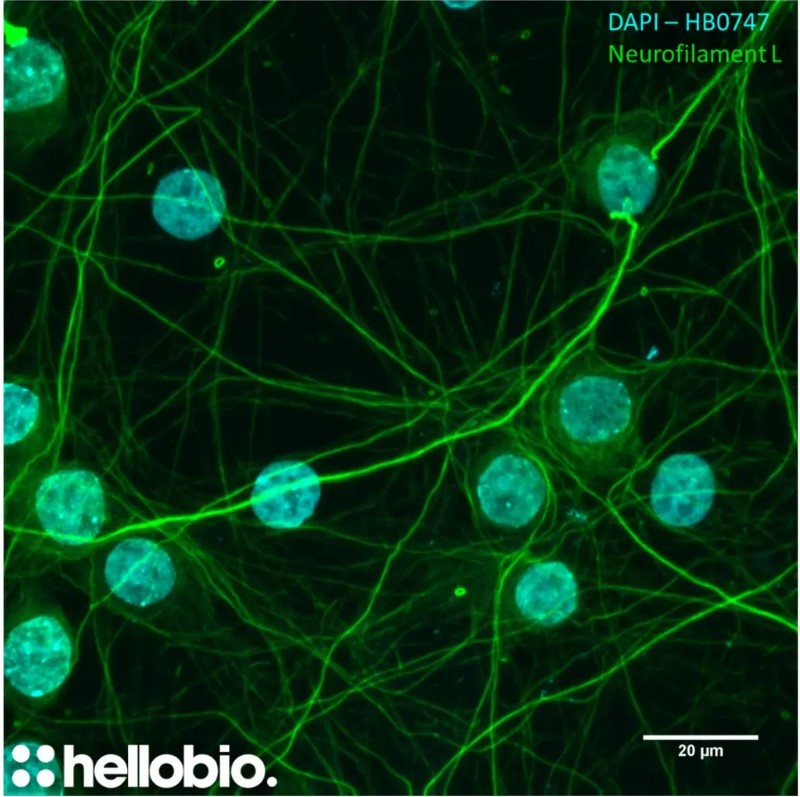
DAPI Staining Solution (1mg/mL) | Hello Bio

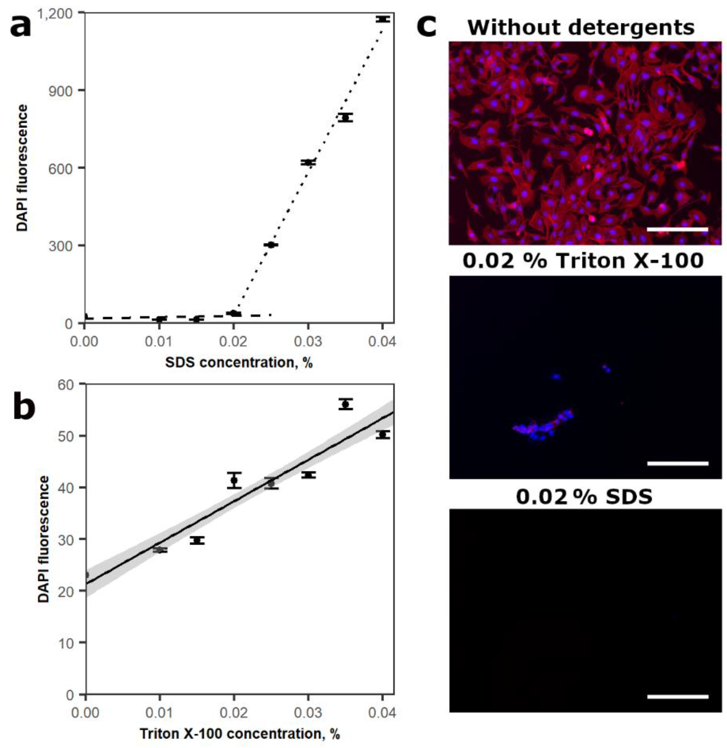
CIMB | Free Full-Text | DNA-DAPI Interaction-Based Method for Cell  Proliferation Rate Evaluation in 3D Structures
CIMB | Free Full-Text | DNA-DAPI Interaction-Based Method for Cell Proliferation Rate Evaluation in 3D Structures

DAPI staining in HCT-116 cells. The cells were treated with test... |  Download Scientific Diagram
DAPI staining in HCT-116 cells. The cells were treated with test... | Download Scientific Diagram

Calcium concentration levels imaged by Fluo-3 AM/DAPI counter staining... |  Download Scientific Diagram
Calcium concentration levels imaged by Fluo-3 AM/DAPI counter staining... | Download Scientific Diagram

DAPI Staining Solution| Abcam中文官网

DNA Labelling Using DAPI: Methodology

DAPI staining and DNA content estimation of nuclei in uncultivable  microbial eukaryotes (Arcellinida and Ciliates) - ScienceDirect
DAPI staining and DNA content estimation of nuclei in uncultivable microbial eukaryotes (Arcellinida and Ciliates) - ScienceDirect

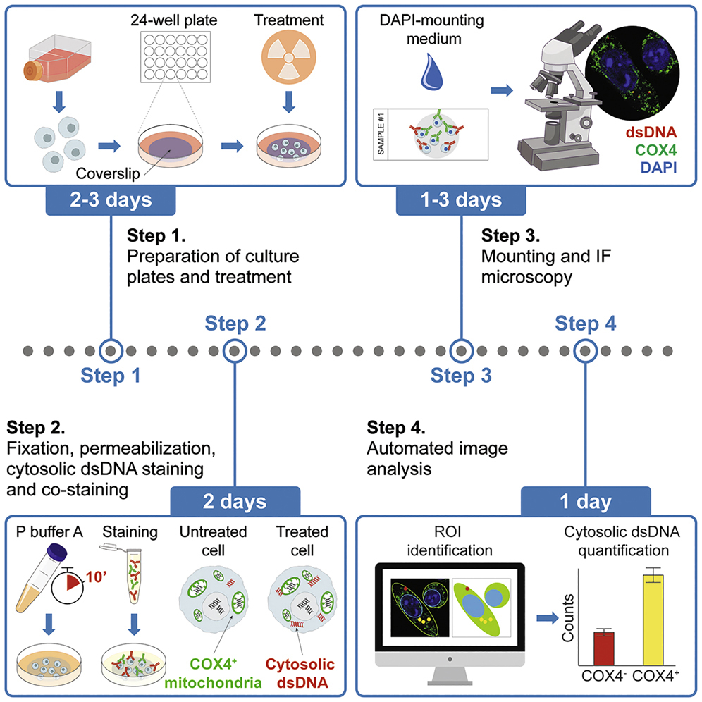
Immunofluorescence microscopy-based assessment of cytosolic DNA  accumulation in mammalian cells
Immunofluorescence microscopy-based assessment of cytosolic DNA accumulation in mammalian cells

Apoptotic nuclear morphological changes highlighted by DAPI staining in  cells treated with graded concentration of NIV and DON (0.5–20 µM) for 24 h.
Apoptotic nuclear morphological changes highlighted by DAPI staining in cells treated with graded concentration of NIV and DON (0.5–20 µM) for 24 h.
Tech Tip: Avoiding Artifacts from UV Photoconversion of DAPI and Hoechst -  Biotium
Tech Tip: Avoiding Artifacts from UV Photoconversion of DAPI and Hoechst - Biotium

Protocol: Staining Cells with Hoechst or DAPI Nuclear Stains - Biotium

DAPI - Wikipedia
DAPI - Wikipedia

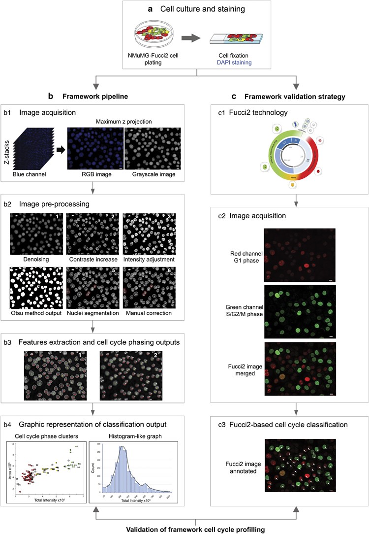
Blue intensity matters for cell cycle profiling in fluorescence DAPI-stained  images | Laboratory Investigation
Blue intensity matters for cell cycle profiling in fluorescence DAPI-stained images | Laboratory Investigation

DAPI and PI double staining of H929 cells. Cell nucleus was visualized... |  Download Scientific Diagram
DAPI and PI double staining of H929 cells. Cell nucleus was visualized... | Download Scientific Diagram

DAPI staining showing 7-Methoxyheptaphylline induces... | Download  Scientific Diagram
DAPI staining showing 7-Methoxyheptaphylline induces... | Download Scientific Diagram